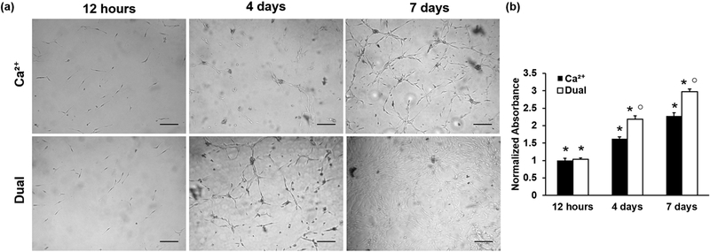
Figure 3.
(a) Photomicrographs of MC3T3-E1 cells seeded on calcium- and dual-crosslinked hydrogels and cultured over 7 days. Top row, Ca2+-crosslinked; bottom row, dual-crosslinked. (Scale bars = 200 μm.) (b) Proliferation of cells seeded on Ca2+ or dual-crosslinked alginate-RGD hydrogels as quantified by MTS assay (*p < 0.01 compared to all other time points for same crosslinking type, ○ p < 0.01 compared to different crosslinking mechanism for the same time point).

