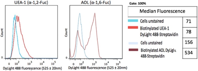
Figure 4.
Cell-surface expression of core and terminal fucose on DU145 cells determined by flow cytometry. DU145 cells were stained with biotinylated lectins, AOL specific for α-1,6, and UEA-1 specific for α-1,2 for Fuc. Stained cells were then treated with DyLight 594 streptavidin. The background control was cells with only DyLight 594 streptavidin staining. Cells were analyzed by Beckman Coulter Cytomics FC500 flow cytometry and CxP software (Beckman Coulter). Overlay histograms are displayed. The median fluorescence for each histogram is assessed for 5×10⁵ acquired cells (100% gated). The data are a representation of one out of two experiments showing similar results.

