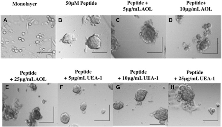
Figure 5.
Phase-contrast images of DU145 MCTS formation on day 5 after incubation with cyclo-RGDfK(TPP) peptide and blockade of core and terminal fucose with AOL and UEA-1 lectins. Phase-contrast images of DU145 cells treated with a combination of 50 µM cyclo-RGDfK(TPP), referred to as “peptide,” and AOL or UEA-1 lectin at concentrations of 5 µg/mL, 10 µg/mL, and 25 µg/mL. Cells at 10,000 were plated per well in a 96-well plate for 5 days. (A) Monolayer of DU145 cell culture, (B) DU145 MCTS formed with peptide only, (C) DU145 MCTS formed with peptide + 5 µg/mL AOL, (D) DU145 MCTS formed with peptide + 10 µg/mL AOL, (E) DU145 MCTS formed with peptide + 25 µg/mL AOL, (F) DU145 MCTS formed with peptide + 5 µg/mL UEA-1, (G) DU145 MCTS formed with peptide + 10 µg/mL UEA-1, and (H) DU145 MCTS formed with peptide + 25 µg/mL UEA-1. The images are representative of two fields of view in two independent experiments. The images were taken at a magnification of 100×. The scale bar in each image represents 100 µm.

