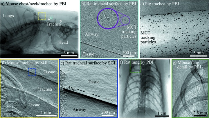
Figure 3.
Typical in vivo X-ray phase-contrast images of the respiratory system. (a) Using propagation-based imaging (PBI), the full length of a mouse trachea from the lungs (left) to the mouth (right) is visible. Coloured boxes are indicative of the approximate imaging locations of the panels shown with corresponding colour. PBI reveals the airway surfaces and mucociliary transit (MCT) tracking particles at high resolution, captured here in (b) a rat and (c) a pig. Alternatively, single-grid imaging (SGI) can be applied to capture an image sensitive to the derivative of the phase, which reveals the curvature of the trachea in (d) a mouse (tiled image), and, at higher magnification (blue box), reveals (e) the airway surface liquid (ASL), shown here in a rat (note the ‘saturated’ phase signal near the ASL/Airway edge has been set to black). (f) PBI enhances the visibility of the lungs, providing (g) a sharp boundary around the edge and a speckle pattern from the air sacs in the lung, shown here in (f) a rat and (g) a mouse. In all cases shown here, the image capture was synchronized to the breath. Note that respiratory images captured in mice and rats are very similar, simply scaled for the animal size. Panel (a) was collected at 25 keV on BL20B2, and panels (b), (d) and (e) on BL20XU at the SPring-8 synchrotron. Panels (c) and (f) were captured at 55 keV on the IMBL at the Australian Synchrotron, and panel (g) was captured at 25 keV at the Munich Compact Light Source (cropped here from the full image to show detail).

