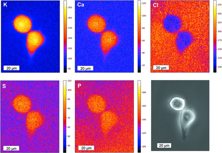
Figure 4.
XRF single element maps (180 × 221 pixels per map) obtained from HeLa cells with corresponding light microscopic image. The scale bar for the XRF maps and the light microscopy image is 20 µm. The color bar to the right of the XRF maps represents the total integrated counts of the detector per pixel during the selected acquisition time. The XRF maps were recorded with a 400 nm pixel size and an exposure time of 0.03 s. The total scanning duration was 30 min.

