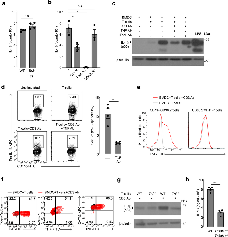
Figure 2. T cell derived TNFα is critical for induction of pro-IL-1β in BMDCs.
(a) lL-1β, as quantified by ELISA, in the supernatants of WT TH0 cells co-cultured with WT or Tlr2/4−/− BMDCs in the presence of CD3 Ab for 6 h. Error bars indicate SEM for n=4 independent experiments. (b) IL-1β measured by ELISA following 6 h of culture with WT TH0 cells and WT BMDCs in the presence of CD3 Ab and neutralizing TNF Ab (20μg/mL), FasL Ab (10μg/mL), or CD40L Ab (20μg/mL). Error bars indicate SEM for n=3 independent experiments. (c) Immunoblot analysis of pro-IL-1β (p35) in the lysates of WT BMDCs stimulated with LPS (100ng/mL) or cultured with TH0 cells in the presence of CD3 Ab and neutralizing antibodies. Data are representative of two independent experiments. (d) Expression of intracellular pro-IL-1β measured by flow cytometry in WT live, CD90-CD11c+ BMDCs cultured with TH0 cells in the presence of CD3 Ab and neutralizing TNF Ab (20μg/mL) for 6h. Flow plots are representative of four independent experiments. Error bars indicate SEM for n=4 independent experiments. (e) Expression of intracellular TNF measured by flow cytometry in WT TH0 cells (left; live,CD11c-CD90.2+) and WT BMDCs (right; live,CD90.2-CD11c+) that were co-cultured for 3 h in the presence of CD3 Ab and brefeldin A. Data are representative of two independent experiments. (f) Expression of intracellular TNF measured by flow cytometry in effector CD4+ T cells (live,CD11c-CD90.2+) polarized to TH1, TH2 and TH17 lineages cultured with WT BMDCs in the presence of CD3 Ab and brefeldin A for 3h. Cells were considered to be transcription factor positive based on isotype control antibody staining. Data are representative of three independent experiments. (g) Immunoblot analysis of pro-IL-1β (p35) in the lysates of WT or Tnf−/− TH0 cells cultured WT BMDCs in the presence or absence of CD3 Ab for 6h. Data are representative of two independent experiments. (h) lL-1β was quantified by ELISA in the supernatants of WT TH0 cells co-cultured with WT or Tnfrsf1a−/−Tnfrsf1b−/− BMDCs in the presence of CD3 Ab for 6 h. Error bars indicate SEM for n=4 independent experiments. (a, b, d, h) Statistical analysis was performed by paired, one-tailed Student’s t-test. *p<0.05, **p<0.01, ***p<0.001, n.s.=not significant.

