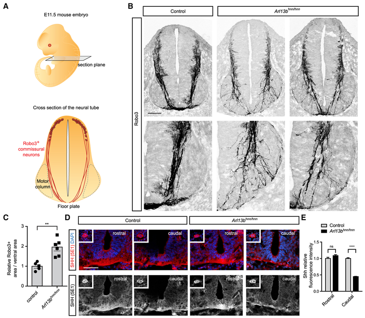
Figure 1. Abnormal Commissural Axon Projections and Midline Crossing in Arl13bhnn/hnn Spinal Cord.
(A) Schematic of an E11.5 embryo and cross-section of the neural tube at the forelimb level. Sections are labeled with Robo3, which marks commissural axons projecting from the dorsal neural tube to the floor plate.
(B) Robo3 immunostaining of E11.5 spinal cord cross-sections of Arl13bhnn/hnn mice.
(C) The area occupied by Robo3+ axons relative to the total area of the ventral neural tube (mean ± SEM) is significantly higher in Arl13bhnn/hnn mice compared to control mice (t test, **p < 0.01).
(D) Shh immunostaining of E11.5 rostral spinal cord cross-sections. Inset shows the notochord.
(E) The Shh relative fluorescence intensity is quantified at both rostral and caudal levels (mean ± SEM). Two-way ANOVA, Bonferroni multiple comparisons (****p < 0.0001). Number of embryos: 5 Arl13bhnn/hnn; 6 control; and 3 sections per embryo.
Scale bars: 100 μm (B) and 50 μm (D).
See also Figure S1.

