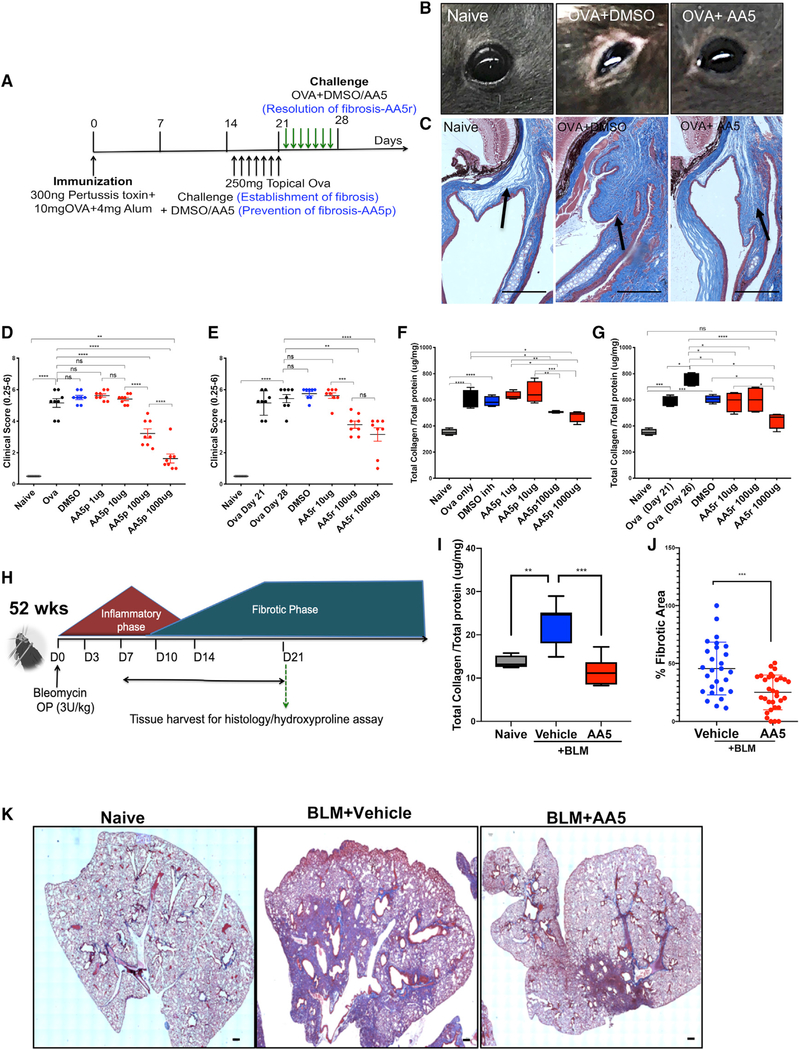
Figure 7. In Vivo Analyses Confirm the Anti-fibrotic Efficacy of the Hit Molecule.
(A) Schema illustrating the experiment to induce and treat ocular fibrosis in mice in a dose-dependent manner.
(B) Representative images of eyes of mice from naive, OVA-sensitized and either DMSO- or AA5-treated animals on day 21.
(C) Representative Gömöri trichrome stained sections of whole eyes (n = 12 per group). Scale bar: 50 μm.
(D and E) Ocular surface inflammatory score in OVA-treated mice (n = 12) treated daily with topical eye drops containing DMSO or (0.1–1,000 μg) AA5 in fibrosis prevention (D) and resolution (E) studies.
(F and G) Total collagen content in conjunctival tissue in naive, OVA-, DMSO-, and AA5-prevention (F) or AA5-resolution (G) treated animals following ocular scarring (n = 12).
(H) Schema illustrating the experiment to induce and treat IPF in mice.
(I) Hydroxyproline content in lung tissue collected on day 21 post -BLM injury with and without AA5 (n = 8).
(J) Percentage fibrotic area measured using the spline contour tool in each sectioned lobe. Data represent mean of two samples analyzed by unpaired t test(***p < 0.001). Related to Figure S7A.
(K) Representative trichrome-stained sections of BLM-treated lungs with and without AA5 collected at day 21 used for scoring fibrotic area in Figure 7J. Scale bar, 200 μm.
Data represent min-max and median. ****p < 0.0001, ***p < 0.001, **p < 0.01, and *p < 0.05 using one-way ANOVA and Sidak’s multiple comparison test.

