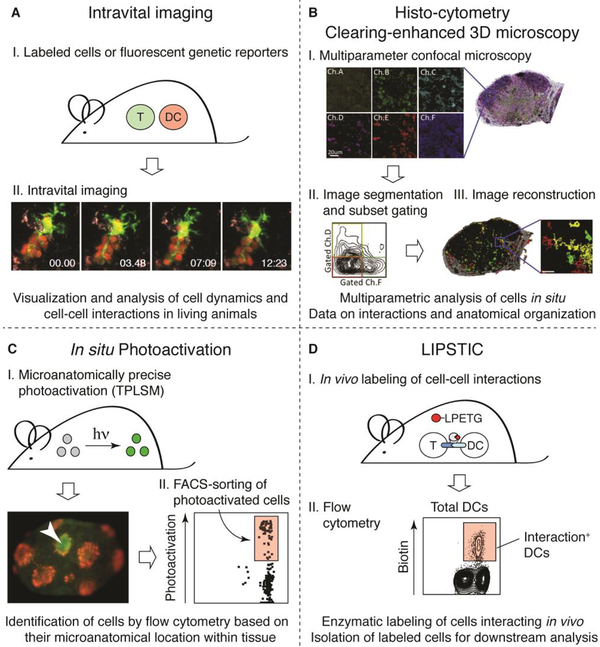
Figure 1. Studying the spatiotemporal dynamics of antigen presentation.
(A) Intravital imaging. (I) Interacting cells are labeled with different fluorescent dyes or by expression of different fluorescent reporter proteins. (II) A two-photon microscope is used to visualize DC-T cell interactions in LN or other locations in explanted tissues or more often within living mice. A cluster of CD8+ T cells (red) undergoing stable interactions with a single antigen-pulsed DC (yellow) is shown (image by G. Victora and K. Swee). (B) Histo-cytometry. Histo-cytometry combines multicolor immunophenotyping with automated image analysis, providing detailed information on the microanatomical location and phenotypic identity of cells in a tissue. (I) A series of confocal images of tissue sections is taken. (II) Images are segmented into cells and, analogously to flow cytometry, channels are compensated for fluorophore spillover. (III) Image analysis allows for quantitative visualization of phenotypically distinct immune cell populations. Images by M. Gerner and R. Germain, adapted with permission from [42]. (C) In situ photoactivation. (I) A microanatomical region of interest is photoactivated within a tissue using MPLSM. An example of photoactivation of cells within a single germinal centers TPLSM is shown (red, follicular dendritic cells; green and arrowhead, photoactivated cells). Image by J. Jacobsen. (II) The tissue is then isolated and dissociated, and photoactivated cells can be easily identified based on their fluorescence in the photoactivation channel by flow cytometry. Cells can be then be used for downstream analyses such as RNA-seq. (D) LIPSTIC. (I) A receptor-ligand pair of interest is genetically tagged with the transpeptidase sortase A (SrtA) or with the SrtA target, 5 N-terminal glycines (G5). The peptide substrate biotin (red)-LPETG is administered by injection, and DCs that interacted with T cells can be isolated based on presence of the biotin-LPETG tag using flow-cytometry. (II) Flow-cytometry contour plot showing biotin-positive cells after in vivo LIPSTIC labeling of DC-T cell interactions via the CD40-CD40L axis.

