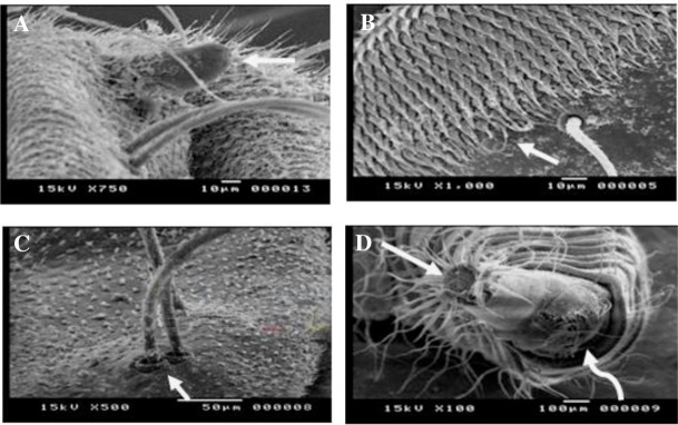
Fig 3. Scanning Electron microscopy of Clogmia albipunctatus body segments and the caudal end.
(A) A nipple shaped anterior spiracle inserted laterally on the anterior annulus of the prothorax, with a solitary pore (white arrow). (B) A dorsal view of thoracic sensory spination with heavily packed tooth-like scales, arranged in several rows, each terminates sharply with a single bristle (white arrow). (C) All setae protrude individually from a hollow basal pocket (white arrow) with small barbs coming off the shaft. (D) The ventral surface of the siphon with the posterior spiracles opening at the apex of the respiratory tube (straight arrow), and a ventrally located anal papilla (curved arrow).

