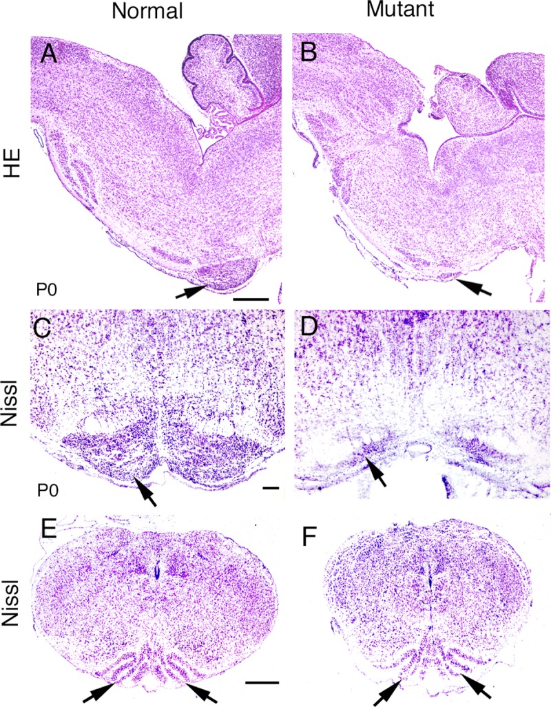
Fig 4. Morphological changes in the hindbrains of Bmpr knockout mutants.

When BMP signaling was eliminated, most of the pontine gray nucleus was lost (A-D), while the morphology of the inferior olivary nucleus appeared unaffected (E, F). (A, B) H&E staining shows that the pontine gray nucleus was dramatically decreased in the Bmpr double knockout mutant (B, arrow) compared to normal (A, arrow). (C, D) Nissl staining of coronal sections shows similar results. Arrows indicate the pontine gray nuclei. (E, F) Arrows indicate the inferior olivary nuclei in both Bmpr double knockout (F) and normal (E) animals. Scale bar: A (for A, B), 500 μm; C (for C,D), 100 μm; E (for E,F), 400 μm.
