Figure 2.

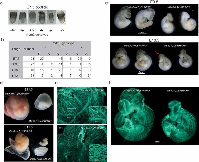
Mdm2Δ7−9/Δ7−9; Trp53RR/RR embryos show severe developmental defects. (a) Representative images of E7.5 embryos demonstrating absence of developmental abnormalities in Mdm2Δ7−9/Δ7−9;Trp53RR/RR embryos at this stage. (b) Phenotype analysis of Trp53RR/RR embryos with different Mdm2 genotypes reveals developmental defects in Mdm2Δ7−9/Δ7−9;Trp53RR/RR embryos starting from E9.5. N – normal, A – abnormal morphology. Asterisk – embryos were partially resorbed. (c) Representative images of Trp53RR/RR embryos with hetero- and homozygous Mdm2 deletion at stages E9.5 (TS15, upper panel) and E10.5–11 (TS18, bottom panel) show progressive growth retardation, abnormal head morphology and neural tube closure defects (arrow) in Mdm2Δ7−9/Δ7−9;Trp53RR/RR embryos. (d) Representative image of Mdm2+/Δ7−9 and Mdm2Δ7−9/Δ7−9;Trp53RR/RR embryos at stage E11.5, (TS19) with the intact (upper panel) and opened (bottom panel) embryonic envelope demonstrates a “bloodless” phenotype of the Mdm2Δ7−9/Δ7−9;Trp53RR/RR embryos. (e) Representative picture of PECAM-1 (endothelial marker) staining in whole mount yolk sac samples shows a reduced number of large blood vessels and decreased branching of the capillary network (zoom-in) in homozygous Mdm2Δ7−9/Δ7−9;Trp53RR/RR embryos (bottom panel). (f) Representative picture of Pecam-1 staining in whole mount embryos shows a reduced branching in blood vessel network and abnormal head morphology of homozygous Mdm2Δ7−9/Δ7−9;Trp53RR/RR embryos.
