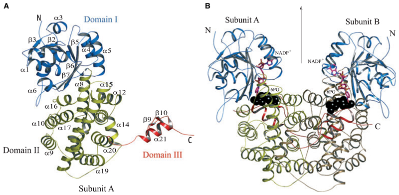
Fig. 3.
(A) Ribbon diagram of an LlPDH subunit. Elements of secondary structure are coloured according to domain as described in Fig. 2 and labelled. The N- and C-termini are marked. (B) The LlPDH dimer viewed perpendicular to the molecular twofold axis of symmetry, which is marked by an arrow. Black spheres depict the position of the substrate (6PG) at the catalytic centre, a stick model is shown for NADP+ and the cofactor is colored according to atom type; C is pink, N is blue, O is orange and P is yellow.

