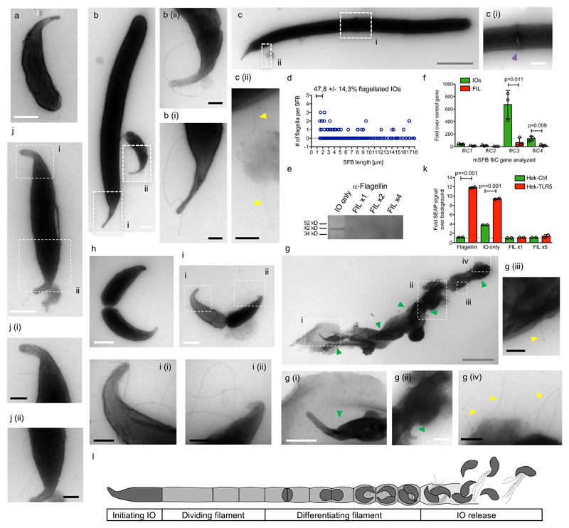
Figure 2. SFB flagellation occurs during in vitro growth and IO development.
a-c. TEM images of in vitro-grown mSFB without (a) and with (b-c) flagella and (c) a septum. d. Summary for in vitro-grown mSFB with mean percent, +/- SE, of 1 to 2 μm-long flagellated SFB that are flagellated. e. Anti-flagellin western blot of IO-only and filament-enriched (FIL) mSFB fractions. The full gel blot from which this panel was generated is available as Supplementary Figure 8c. f. qPCR analysis of fliC genes of in vivo-grown mSFB fractions. TEM images of (g) in vitro-grown mSFB with flagella in the vicinity of IOs released from a ruptured filament (n=2) and (h-j) in vivo-grown mSFB without (h) and with (i-j) flagella during IO development (n=5 and n=8, respectively). k. TLR5 stimulation by mSFB fractions using HEK reporter cell lines. l. Schematic overview of the SFB replicative life-cycle. Arrows: yellow: flagella; green: IOs. In (e/k), “x” denotes fold increase in SFB numbers assayed. Scale bars: white: 500 nm; black: 200 nm; grey: 2 μm. (e, f, k) Data from one of two (e), from three (f), and one of three (k) independent experiments with similar results. (f/k) Mean +/- standard deviation and two-sided t-test statistical analysis. TEM images are from four in vitro (n=278) and four in vivo (n=320) independent mSFB experiments with similar results.

