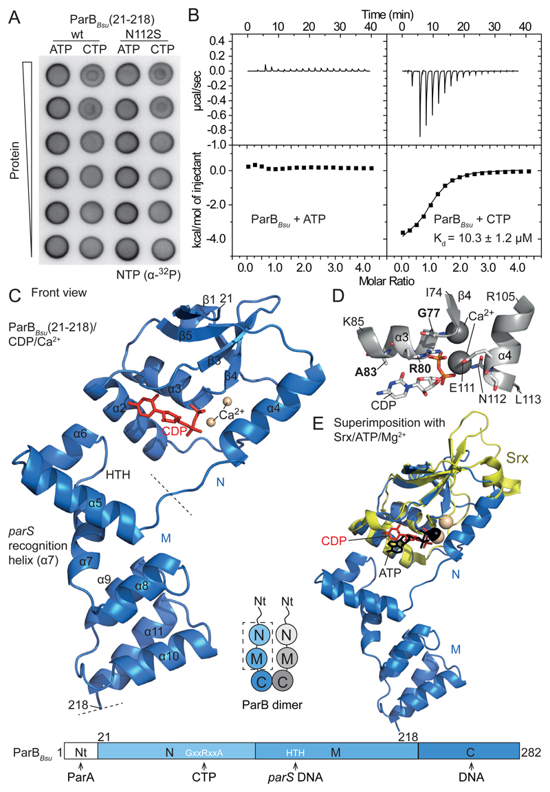
Fig. 1. ParB CTP binding.
(A) Membrane-spotting assay using radiolabeled nucleotides. (B) ITC measurements with full-length ParBBsu in the presence of Ca2+. The Kd obtained in a typical experiment is given. The interval indicates deviations of data points from the fit. (C) Crystal structure of a single chain of ParBBsu(21-218)/with CDP/Ca2+. (D) Nucleotide binding pocket in ParBBsu/CDP/Ca2+. GxxRxxA residues marked in bold. (E) Superimposition of ParBBsu(21-218)/CDP/Ca2+ with human Srx/ATP/Mg2+ (PDB: 3CYI).

