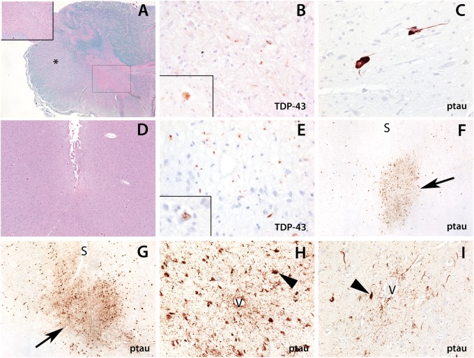
Figure 1.
Neuropathological features of ALS with chronic traumatic encephalopathy in CTE-5 (A–F) and CTE-8 (G–I). (A–C) There is marked degeneration of the spinal cord. (A) Luxol H&E shows degeneration of the lateral corticospinal tracts (*, ×20 magnification) and loss of anterior horn cells (box and inset, ×200 magnification). (B) An immunostain for phosphorylated TDP-43 (pTDP-43) shows rare cytoplasmic inclusions within the remaining anterior horn cells (×200 and inset, ×400 magnification). (C) Immunostaining (AT8) shows scattered ptau-positive neurofibrillary tangles and threads within the anterior horn (×400 magnification). (D–F) The cortex shows the changes of chronic traumatic encephalopathy. (E) There are pTDP-43-positive neurites and cytoplasmic inclusions that are predominant within the sulcal depths (×200 magnification) and occasional skein-like inclusions within neurons (inset, ×400 magnification). (F, G) Ptau pathology is present focally at the sulcal depths of the frontal cortex with perivascular neurofibrillary tangles and threads in CTE-5 (F) and CTE-8 (G) ([S], sulcus; arrows highlight the foci of ptau pathology; ×40 magnification). (H, I) Perivascular ptau neurofibrillary tangles and processes in CTE-8 ([V], blood vessel; arrowheads highlight neurofibrillary tangles; ×200 magnification).

