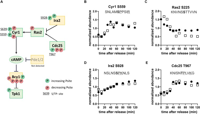
FIGURE 6.
The protein kinase A pathway is phospho-regulated through the cell cycle. (A) Map of the Ras-branch of the PKA pathway. Circles indicate sites whose phosphorylation increases (green, clusters 1–3) or decreases (red, clusters 4–5) through the cell cycle. Only sites found in both replicates are reported. S/TP sites, possibly phosphorylated by cyclin-dependent kinases, are denoted by their residue numbers adjacent to the phosphorylation site. (B–E) Examples of dynamic phosphorylation of sites on different upstream regulators of PKA through the cell cycle. Residues associated with consensus cyclin-dependent kinase sites are underlined and the phosphorylated residue is shown in bold. Black and white symbols denote the two replicate time courses.

