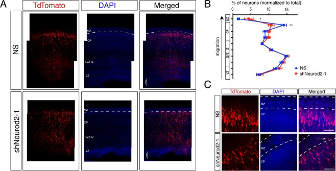
Figure 6.
NEUROD2’s role in neuronal soma positioning to the primitive cortical zone. (A) Embryos (at E14.5) were in utero electroporated with tdTomato fluorescent marker and with NS (non-silencing) shRNA or with shNeurod2-1. Embryos were retrieved at E17.5, coronally sectioned, and tdTomato signal was subsequently imaged by confocal microscopy. VZ (ventricular zone), SVZ-IZ (subventricular-intermediate zone), CP (cortical plate), and MZ (marginal zone) are labeled. Scale bar: 100 µm. (B) Transfected neurons (a total of n = 16,184 for NS and n = 19,339 for shNeurod2-1) derived from in utero electroporation of seven littermate embryo pairs from five independent pregnant mice were counted. Normalized percentages of tdTomato-positive neurons counted in individual zones are plotted. Significantly less neurons are localized to the upper cortical plate in neurons where Neurod2 is knocked down. (C) Higher magnification images of the MZ and the CP in NS and shNeurod2-1 electroporated cortices. White arrow points to occasional neurons that over-migrate into the MZ of the shNeurod2-1 transfected samples. Scale bar: 50 µm. Bars represent S.E.M. p < 0.0001 by two-way ANOVA and *p = 0.0165 and **p = 0.0015 by post hoc Sidak’s test.

