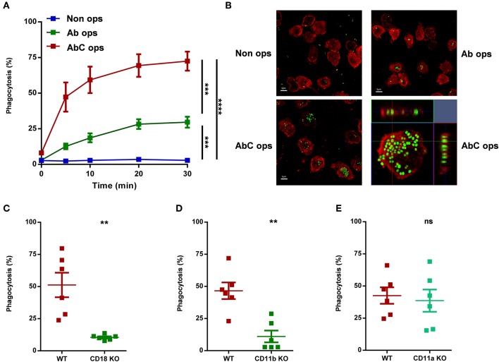
Figure 1.
Participation of different receptors in phagocytosis of human (A,B) or murine (C–E) neutrophils. (A) Analysis of phagocytosis of non-opsonized, partially, or completely opsonized GFP expressing S. aureus by human PMN. Kinetics of phagocytosis ± SEM, n = 4. Data were compared after 30 min phagocytosis using RM-ANOVA coupled with Tukey's post hoc test. (B) Confocal microscopic images of human neutrophils after 20 min phagocytosis of non-opsonized (UL), partially (UR), and completely (LL) opsonized GFP expressing S. aureus; red color shows CD11b labeling. X-Y projections of engulfed bacteria with the respective side views (LR). Representative images out of 4 independent experiments. (C–E) Quantification of phagocytosis of WT vs. CD18 KO (C), CD11b KO (D), CD11a KO (E) murine PMN. Data were compared using Student's t-test; n = 6, 6, 6 ± SEM. **P < 0.01; ***P < 0.001; ****P < 0.0001.

