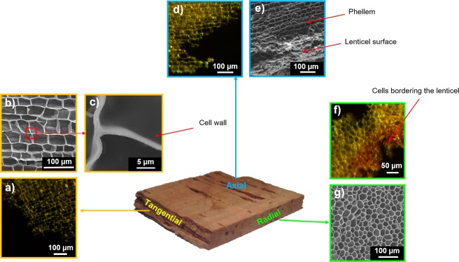
Figure 4.
Cork structure observed from optical microscopy (OM) and scanning electron microscopy (SEM). View from the: Tangential direction (or radial plane). (a) Phellem, from OM. b) Phellem, from SEM. (c) Zoom in the cork cell wall. Axial direction (or transverse plane). (d) Phellem, from OM. (e) Phellem and lenticel surface, from SEM. Radial direction (or tangential plane). (f) Phellem and lenticel, from OM. (g) Phellem, from SEM.

