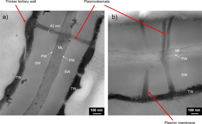
Figure 6.
Phellem cell walls observed from Transmission Electron Microscopy (TEM). The cell wall is composed of the four successive layers: Middle Lamella (ML), Primary Wall (PW), Secondary Wall (SW) and Tertiary Wall (TW). The selected TEM pictures highlight a plasmodesmata crossing the cell wall. (a) A thicker region of the tertiary wall can be seen at the extremities of the plasmodesmata. (b) The plasmic membrane is visible inside the plasmodesmata.

