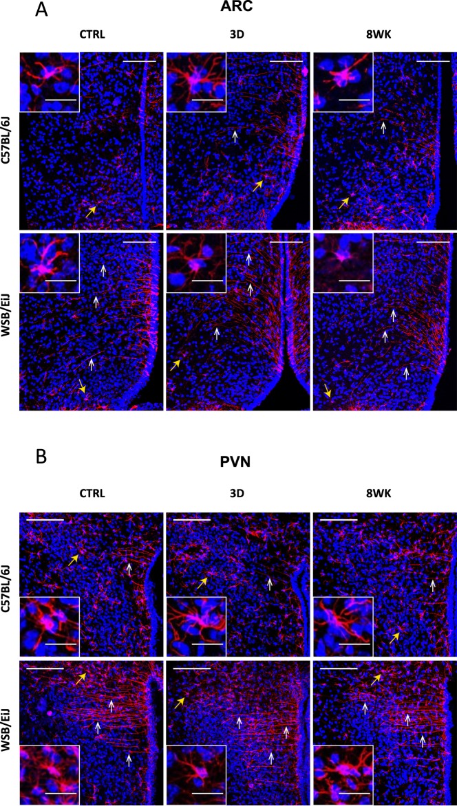
Figure 6.
WSB/EiJ mice show remarkably well-defined elongated ependymal cells in both the hypothalamic arcuate nucleus (ARC) and the hypothalamic paraventricular nucleus (PVN), whatever the diet is. Immuno-histochemical analysis of GFAP-positive cells (glial cells including astrocytes and tanycytes) in the hypothalamic nuclei of twelve week-old C57BL/6J and WSB/EiJ male mice either maintained under control feeding (CTRL), or challenged with HFD for the last 3 days of the period (3D) or during the whole 8-week experiment duration (8WK). (A) Representative confocal images of GFAP-positive cells (astrocyte- and tanycyte-like glial cells; in red) and DAPI-stained nuclei (in blue) in the ARC region of coronal sections of C57BL/6J (top panel) and WSB/EiJ (bottom panel) mice. GFAP-positive elongated tanycyte-like ependymal cells (pointed by white arrowheads) were present in the top region of ARC close to the third ventricle (3 v) at a much greater extent in WSB/EiJ sections than in C57BL/6J. In contrast, C57BL/6J sections showed a greater proportion of shorter and well distinguishable GFAP-positive astrocyte-like cells (pointed by yellow arrowheads; magnified in insets). (B) Representative confocal images of GFAP-positive cells (in red) and DAPI-stained nuclei (in blue) in the PVN region of coronal sections of C57BL/6J (top panel) and WSB/EiJ (bottom panel) mice. The effect of strain on the morphology of GFAP-positive cells (mostly astrocyte-like cells (yellow arrowheads) in C57BL/6J mice vs mostly elongated ependymal cells (white arrowheads) in WSB/EiJ sections) is even more pronounced in the PVN region than in the ARC. Scale bars in main images: 100 µm; scale bars in insets: 20 µm.

