
Figure 6. AgLDL-stimulated Akt activation regulates actin polymerization and lysosome exocytosis at the LS and occurs in a PI3K/SYK dependent manner.
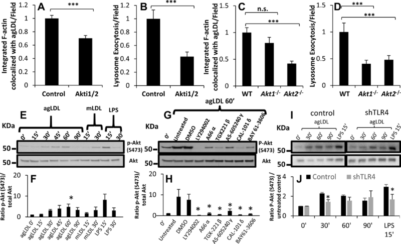
(A) Actin polymerization at the LS was analyzed by confocal microscopy of J774 cells pre-treated with DMSO (control) or Akt inhibitor Akti1/2 (4 μM) for 1 hr prior to treatment with A546-agLDL for 1 hr in the presence or absence of inhibitor. Cells were fixed, and F-actin stained using A488-phalloidin, prior to analysis. Confocal images were used to quantify F-actin colocalized with agLDL per field. (B) Lysosome exocytosis at the LS was analyzed by confocal microscopy. J774 cells were incubated with biotin-fluorescein-dextran overnight and chased in media to dextran load their lysosomes. Cells were pre-treated with the same inhibitors as in (A) prior to treatment with Streptavidin-A546-agLDL for 90 min in the presence or absence of inhibitors. Cells were incubated with an excess of biotin, fixed, permeabilized with triton X-100, washed extensively and analyzed. Confocal images were used to quantify exocytosed biotin-fluorescein-dextran colocalized with Streptavidin-A546-agLDL (lysosome exocytosis) per field. (C) Actin polymerization at the LS was analyzed by confocal microscopy of WT, Akt1−/− and Akt2−/− BMM treated with A546-agLDL for 1 hr and quantified per field. (D) Lysosome exocytosis at the LS was assessed by confocal microscopy of WT, Akt1−/− and Akt2−/− BMM treated with Streptavidin-A546-agLDL for 90 min and quantified per field. (E) J774 cells were treated with agLDL (1 mg/mL), mLDL (1 mg/mL) or LPS (100 ng/mL) for indicated periods of time, prior to lysis and immunoblot analysis. (F) Ratios of phosphorylated (S473) to total Akt protein were obtained by densitometry analysis. (G) J774 cells were pre-treated for 1 hr with various inhibitors (LY294002 50 μM, A66 8 μM, TGX-221 2 μM, AS-605240 2 μM, CAL-101 2 μM, and BAY 61–3606 5 μM), left untreated or treated with DMSO control, and subsequently incubated with agLDL (1 mg/mL) for 1 hr in the presence or absence of inhibitors prior to lysis and immunoblot analysis. (H) Ratios of phosphorylated (S473) to total Akt protein were obtained by densitometry analysis. (I) J774 cells stably transfected with control shRNA or TLR4 specific shRNA (shTLR4) were treated with agLDL (1 mg/mL) for indicated periods of time prior to lysis and immunoblot analysis. (J) Ratios of phosphorylated (S473) to total Akt protein were obtained by densitometry analysis.. Data were compiled from 3 independent experiments. Error bars represent the SEM. * p < 0.05, ** < 0.01, *** p ≤ 0.001. n.s. not statistically significant.