
Figure 4. Viral replication compartments are reorganized during the late stage of infection.
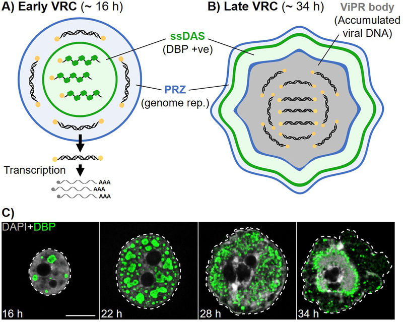
A. Schematic representation of an early viral replication compartment (VRC), showing the single-stranded DNA accumulation site (ssDAS) in which ssDNA replication intermediates bound by the viral DNA-binding protein (DBP) are present (green), and the peripheral replicative zone (PRZ) where viral DNA replication takes place (blue). Viral dsDNA genomes move away from VRCs where transcription and RNA processing take place. B. Schematic representation of a late VRC including virus-induced post-replicative (ViPR) body. C. Human bronchial epithelial cells infected with Ad5 showing VRC morphology at 16, 22, 28 or 34 hours post-infection (h). Nuclei were visualized by confocal microscopy with DBP immuno-labelled (green), and DNA labelled with DAPI (grey). Dashed lines outline nuclei. Scale bar = 10 μm. Note, at 34 h VRCs can be observed as a ring surrounding a single large ViPR body.