Fig. 1.
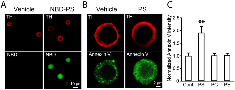
Exogenous PS addition in culture media increased cellular PS in chromaffin cells A. Images of NBD-PS in mouse chromaffin cells. Images of chromaffin cells treated with vehicle (Left) or NBD-PS (Right). Chromaffin cells were stained with anti-tyrosine hydroxylase (Upper). B. Intensity of Annexin V-FITC signal was increased in PS-incubated cells (Right) alternative to control cells (Left). Chromaffin cells were identified with anti-TH antibody (Upper). C. Normalized intensities of Annexin V-FITC signal indicate increased cellular PS level in PS-treated cells (Control: 1.00 ± 0.11, n = 40 cells, PS: 1.92 ± 0.25, n = 32 cells, PC: 1.01 ± 0.07, n = 36 cells, PE: 1.03 ± 0.07, n = 38 cells; one-way ANOVA followed by Turkey’s post hoc test, F(3, 142) = 10.5436, *** p < 0.001, PS vs. Control: ** p = 0.0010, PC vs. Control: p = 0.8999, PE vs. Control: p = 0.8999). Data was pooled from 3 independent cultures.

