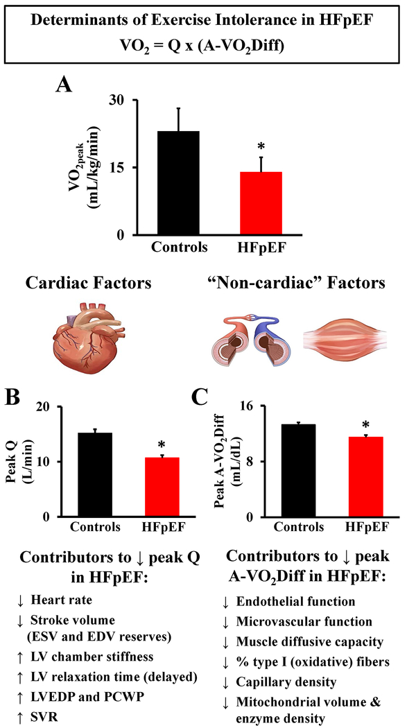
Figure 1.
Magnitude and pathophysiology of exercise intolerance in patients with heart failure and preserved ejection fraction (HFpEF). A. HFpEF patients demonstrate severe exercise intolerance, measured objectively as a ~40% reduction in peak oxygen uptake (VO2peak) (mL/kg/min) during peak aerobic exercise compared to healthy age-matched controls, adapted and pooled (mean ± SD) from published data by Bhella et al. (2011)50, Dhakal et al. (2015)16, and Haykowsky et al. (2011)17. B. HFpEF patients demonstrate reduced peak exercise cardiac output (Q) (L/min), adapted from published data (mean ± SE) by Dhakal et al. (2015)16. C. HFpEF patients demonstrate reduced peak exercise arteriovenous oxygen difference (a-vO2Diff) (mL/dL), adapted from published data (mean ± SE) by Dhakal et al. (2015)16. EDV: end-diastolic volume, ESV: end-systolic volume, LV: left ventricle, LVEDP: left ventricle end-diastolic pressure, SVR: systemic vascular resistance, PCWP: pulmonary capillary wedge pressure. * indicates significant (P <.05) difference between HFpEF and healthy age-matched controls for all figures.

