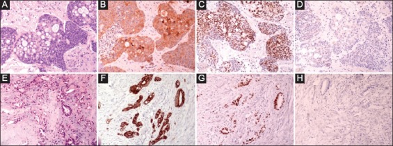
Figure 3.

(A-D) Tissue sections from brain metastasis stained for (A) H&E, (B) CK20, (C) CDX-2 and (D) CK7. E-H: Representative sections (H&E, E) from surgical specimen of mastectomy showed histopathologic characteristics of a moderately differentiated adenocarcinoma with the following immunophenotype: (F) CK20+, (G) CDX-2+ and (H) CK7. The immunophenotype of both cases was consistent with metastatic colorectal carcinoma (magnification ×200)
