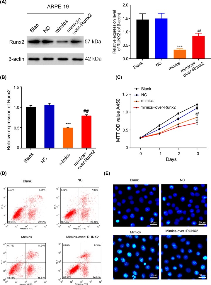
Figure 5. MiR-218 suppressed the proliferation and induced the apoptosis of RPEs via Runx2.
ARPE-19 cells were either transfected with miR-218 mimics or co-transfected with miR-218 mimics or the Runx2 plasmid. (A) Western blot analysis of Runx2 expression; β-actin served as an internal control. (B) RT-qPCR analysis of Runx2 levels, ***P < 0.001 vs. NC group; ##P < 0.01 vs. mimics group. (C) Cell proliferation was examined by the CCK-8 assay, ***P < 0.001 vs. NC group; ##P < 0.01 vs. mimics group. (D) Cell apoptosis was assessed by flow cytometry. (E) Hoechst staining was performed to evaluate cell apoptosis (original magnification ×200, scale bar = 50 μm). All experiments were repeated three times.

