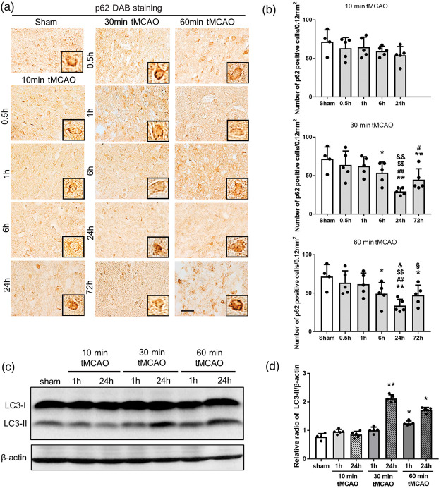
Figure 4.
(a) Immunohistochemical staining of p62 in the peri-ischemic lesion, with (b) quantitative analysis. (c) Western blot for LC3-I and LC3-II levels. (d) Quantitation of LC3-II/β-actin ratio (*p < 0.05 versus sham, **p < 0.01 versus sham; #p < 0.05 versus 0.5 h, ##p < 0.01 versus 0.5 h; $p < 0.05 versus 1 h, $$p < 0.01 versus 1 h; &p < 0.05 versus 6 h, &&p < 0.01 versus 6 h; §p < 0.01 versus 24 h. Scale bar = 50 µm).

