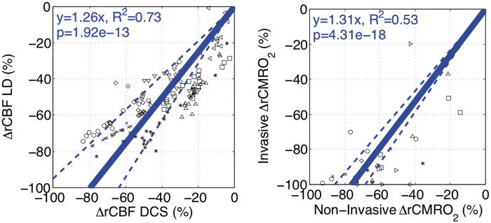
Figure 6.
Validation of non-invasive diffuse correlation spectroscopy (DCS, left) and CMRO2 (right) – data from individual subjects are indicated by a unique symbol. Measurements of change in relative cerebral blood flow using laser Doppler (rCBF LD, %) and DCS (rCBF DCS, %) are compared using a linear mixed-effects model (n = 8; left). DCS measurements demonstrate a good linear correlation (fixed slope effect p < 0.001; R2=0.73) against laser Doppler measurements. Similarly, invasive and non-invasive measurements of rCMRO2 (%) are compared using a linear mixed-effects model (n = 7; right). Non-invasive rCMRO2 quantification demonstrates a significant association but limited linearity (fixed slope effect p < 0.001; R2=0.53) against invasive sampling. Fitted linear relationships (solid line) with 95% confidence intervals (dotted line) are plotted in blue.

