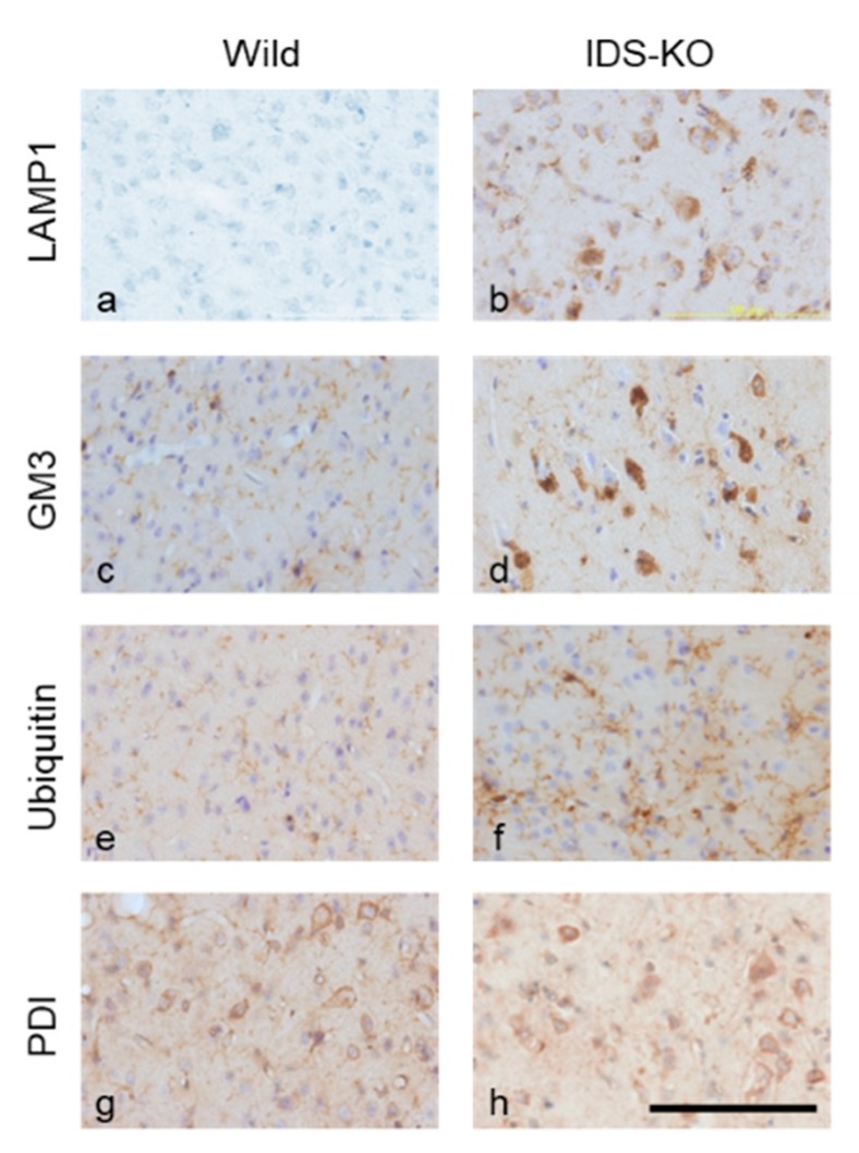
Figure 5.
Immunostaining of the cerebral cortex for lysosomal associated protein 1 (LAMP 1; a,b), GM3 (c,d), ubiquitin (e,f), and protein disulfide isomerase (PDI; g,h). Panels a, c, e, and g were obtained from wild mice, and b, d, f, and h were from IDS-KO mice. A number of cells in the IDS-KO mouse brain were immunopositive for LAMP1, GM3, and ubiquitin; this was not observed in wild-type mice. Both groups had PDI-positive cells, but there was no significant difference in the number. Bar = 100 μm.

