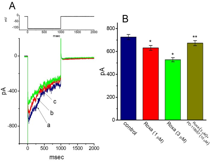
Figure 7.
Effect of roxadustat on erg-mediated K+ current (IK(erg)) in GH3 cells. In this set of experiments, cells were bathed in high-K+, Ca2+-free solution and the recording pipette was filled with K+-containing solution. (A) Representative IK(erg) traces obtained in the absence (a) and presence of 1 μM roxadustat (b) or 3 μM roxadustat (c). The upper part is the voltage protocol used for elicitation of deactivating IK(erg). (B) Summary bar graph showing the effects of roxadustat (Roxa) and roxadustat plus PD-118057 on IK(erg) amplitude in GH3 cells. To evoke deactivating IK(erg), each cell was maintained at -10 mV and the hyperpolarizing pulse to -110 mV with a duration of 1 sec was applied. IK(erg) amplitude was measured at the beginning of hyperpolarizing pulse. Each bar indicates the mean ± SEM (n = 8 for each bar). * Significantly different from control (p < 0.05) and ** significantly different from 3 μM roxadustat alone group.

