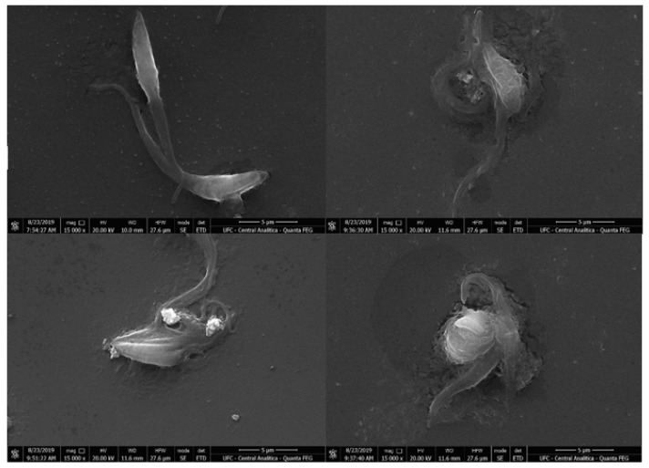
Figure 3.
Scanning electron microscopy images of Trypanosoma cruzi epimastigotes. Untreated epimastigotes (control; (A)), epimastigotes treated with IC50 (B,C) and 2 × IC50 for compound 7 (D). Treated parasites showed ultrastructural changes, such as changes in typical shape, apparent leakage of cytoplasmic content, and cell membrane degradation. Scale bar =5 μm.

