Figure 1.
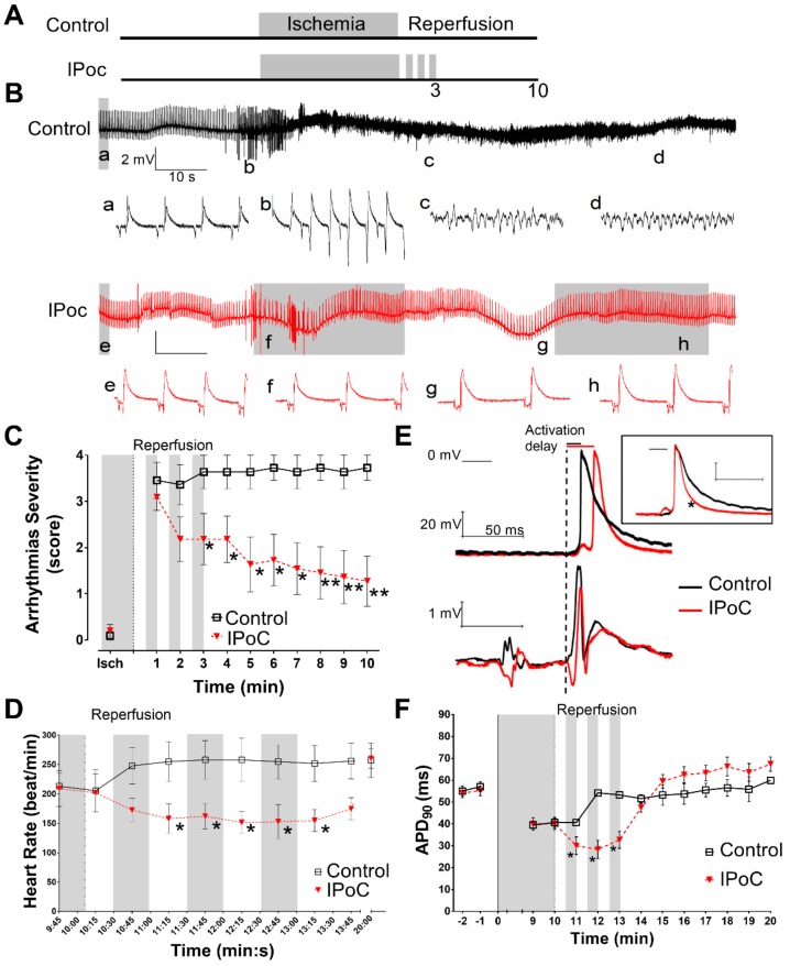
Electrophysiological effects of IPoC in isolated rat hearts. (A) Experimental protocol: 10 min of regional myocardial ischemia (indicated in grey) followed by 10 min reperfusion as Control; IPoC by 3 cycles of reperfusion/regional ischemia, 30 s each. (B) Representatives ECG from the first 2 min of reperfusion. The Control heart developed ventricular fibrillation and IPoC suffered transient episodes of ventricular tachycardia and bradycardia. Lower case letters from a to h corresponds to 1 s traces showed below. (C) The hearts did not develop sustained arrhythmias prior reperfusion. Control group presented severe ventricular arrhythmias through reperfusion whereas IPoC progressively reduced the severity. (D) IPoC induced transient bradycardia. (E) Representative transmembrane potential and ECG simultaneously obtained during the 2nd min of reperfusion. Dashed vertical line indicates the beginning of the QRS complex used to measure the delay to epicardial activation. In the inset, the action potentials were artificially aligned to 0 phase for better identification of action potential duration (APD) shortening. (F) Both groups have similar duration prior to reperfusion reaching values around 40 ms at the end of ischemia. During reperfusion, Control hearts recovered preischemic APD90 values. IPoC induced a transient shortening during the first 3 min of reperfusion. * p < 0.05 and ** p < 0.01 for Control vs. IPoC by repeated measures ANOVA.

