Figure 6.
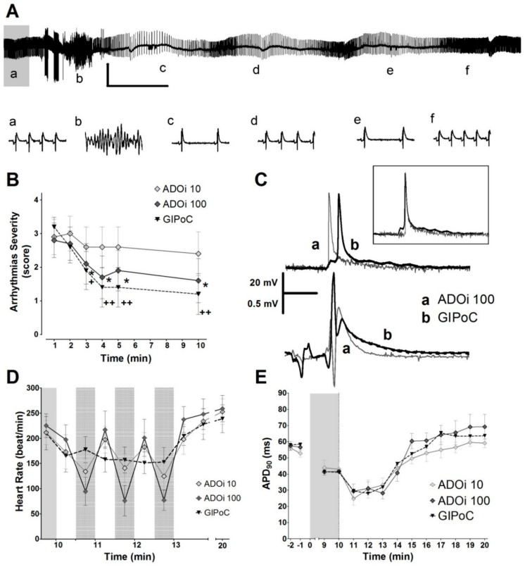
Antiarrhythmic effect and transient bradycardia induced by adenosine and GIPoC. (A) Representatives ECG traces from the first 3 min of reperfusion that show sinus rhythm recovery by intermittent adenosine in a heart that developed a ventricular arrhythmia. Adenosine also induced transient episodes of bradycardia. Vertical scales indicate 1 mV and horizontal 20 s. Lower case letters from a to f indicate the moment in which the corresponding 1 s traces showed below were taken. (B) The higher dose of adenosine almost reduced the severity of arrhythmias to the same level of protection than GIPoC, but the lower dose failed to protect. (C) Adenosine and GIPoC shortened action potential duration, but only GIPoC delayed the activation of the epicardial tissue previously submitted to regional ischemia. Adenosine shortened QT duration. (D) Adenosine and GIPoC induced transient bradycardia, followed by a progressive hear rate recovery towards the minute 10th of reperfusion. Bradycardia was more pronounced in adenosine treated hearts. Grey areas with black dots indicate global ischemia or adenosine treatment. (E) During the first 3 min of reperfusion, ADOi 10, ADOi 100 and GIPoC shortened the action potential duration, and then recover preischemic values. * p < 0.05 and ** p < 0.01 for Control vs. IPoC by repeated measures two-way ANOVA.

