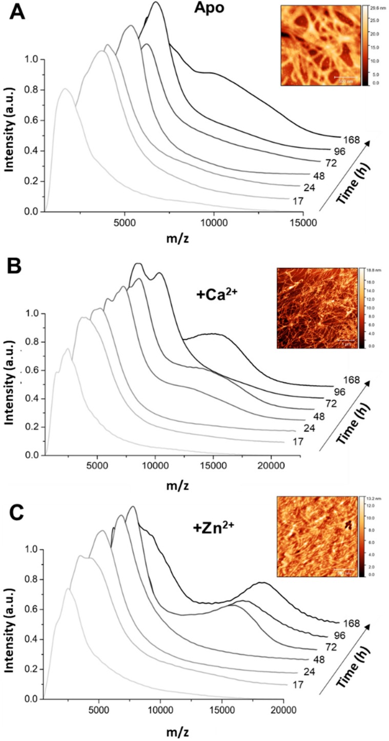
Figure 4.
Different types of aggregates populate Tau aggregation time-points in different conditions. Native mass spectrum of Tau protein in near native state at pH 7.2 shows the formation of Tau aggregates during incubation at 37 °C with agitation (800 rpm). Samples were analysed at seven different time-points (0 h until 168 h corresponding from light grey to black). Aggregation of 35 µM Tau at pH 7.4 with 1 mM DTT, 0.5 mg/mL heparin sodium salt, 50 mM NaCl, 1 mM phenylmethanesulfonyl fluoride (PMSF) in the absence (A) and in presence of 1.1 mM CaCl2 (B) or ZnCl2 (C). Insets: Atomic Force Microscopy (AFM) images of Tau species at 50 h; scale bars: 500 nm in A and C, 1 μm in B.

