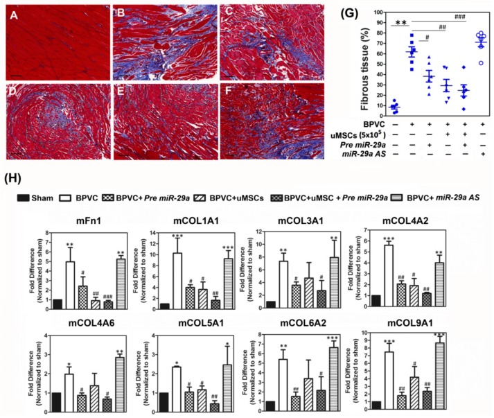
Figure 5.
Administration of miR-29a or transplantation of uMSCs exhibited the anti-fibrotic properties of C57BL/6 mice in vivo and alleviated BPVC-induced gastrocnemius muscle fibrosis. (A–F) Representative immunohistochemistry figures of gastrocnemius muscles with the nuclei stained black, the muscle cells stained red, and fibrous tissue stained blue by Masson’s trichrome staining, after 3 weeks of (A) saline-injection, (B) BPVC-injection, (C) Pre miR-29a administration, (D) uMSCs transplantation, (E) combination of Pre miR-29a and uMSCs treatments, or (F) BPVC + miR-29a AS administration. At least five different tissue sections randomly selected from each group of mice were estimated and their expression patterns were similar (n = 6/each group). Scale bar: 100 µm. (G) After 21 days of uMSCs transplantation, the amount of fibrotic tissues was scored by Masson’s trichrome staining. The level of fibrosis was significantly abridged post 21 day–s of Pre miR-29a administration or uMSCs transplantation, and shown maximal reduced effect by combination of Pre miR-29a and uMSCs treatments. (H) Tissue extracts were collected from gastrocnemius muscles on day 21 post saline or BPVC injections, or after three weeks of Pre miR-29a, miR-29a AS as well as uMSC transplantation post 72 h of BPVC-injection. The protein levels of extracellular matrix (ECM) genes in each group of mice were estimated by using the Quantibody Mouse ECM Array (n = 6/group). Data were shown in means ± SD; * p < 0.05, ** p < 0.01, *** p < 0.001 for the BPVC group vs. the Sham group; # p < 0.05, ## p < 0.01, and ### p < 0.001 for the BPVC group vs. the Sham group; * p < 0.05, and ** p < 0.01 for the BPVC + Pre miR-29a group, the BPVC + miR-29a AS group, the BPVC + uMSC group, the BPVC + uMSC + Pre miR-29a group vs. the BPVC group.

