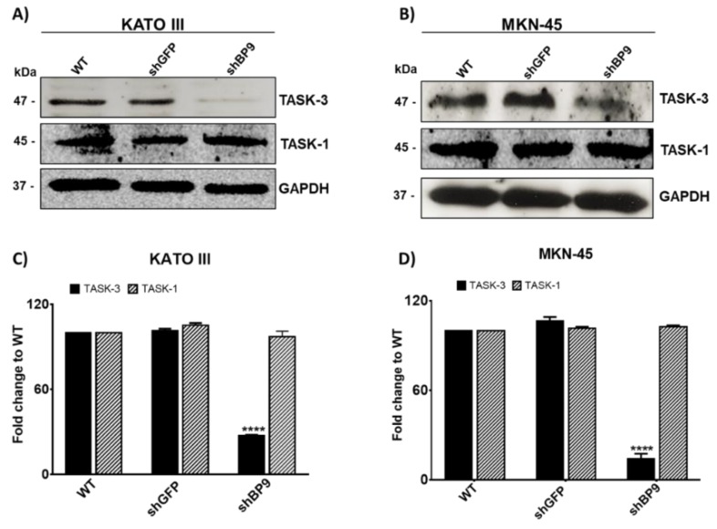
Figure 2.
Protein levels of TASK-3 and TASK-1 in KATO III and MKN-45 cell lines. (A,B) Representative immunoblots for TASK-3, TASK-1, and GAPDH are shown for wild-type (WT) cells as well as cells transduced with shRNAs against GFP (shGFP) or TASK-3 (shBP9). (C,D) Relative abundance of TASK-3 and TASK-1 protein based on densitometric analyses. Data are expressed as mean ± SEM of three independent experiments. **** p < 0.0001, compared with WT, based on ANOVA followed by Dunnett’s test.

