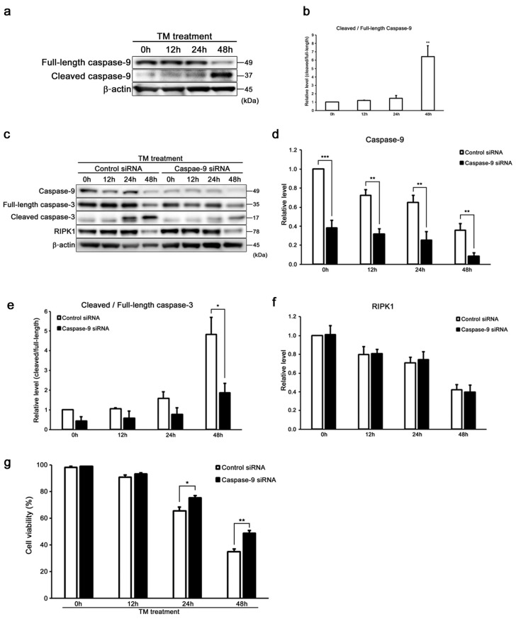
Figure 5.
Caspase-9 influenced the induction of intrinsic apoptosis along with caspase-3 in HEI-OC1 cells. (a,b) Representative Western blots showing the expression of cleaved/full-length caspase-9. β-actin was included as the loading control. The expression of cleaved/full-length caspase-9 was detected, and the means ± S.D. (fold changes compared to the control group) of three or more independent studies were presented (** p < 0.01 compared to the 0 h group, determined using one-way ANOVA followed by Bonferroni test). Full-length blots are presented in Figure S5a–c. (c–f) Representative Western blots showing the expressions of caspase-9, cleaved/full-length caspase-3, and RIPK1 in tunicamycin-treated caspase-9 KD cells (50 µg/mL for 48 h). β-actin was included as the loading control. The expressions of caspase-9, cleaved/full-length caspase-3, and RIPK1 were detected, and the means ± S.D. (fold changes compared to the control group) of three or more independent studies were presented (* p < 0.05, ** p < 0.01, and *** p < 0.001 compared to the control group, determined using unpaired Student’s t-test). Full-length blots are presented in Figure S5d–h. (g) After transfection with caspase-9 and control siRNA for 48 h, the cells were treated with tunicamycin (50 µg/mL for 48 h), and cell viability was determined by trypan blue staining. The data are represented as means ± S.D. of three or more independent studies (* p < 0.05 and ** p < 0.01 compared to the control group, determined using unpaired Student’s t-test).

