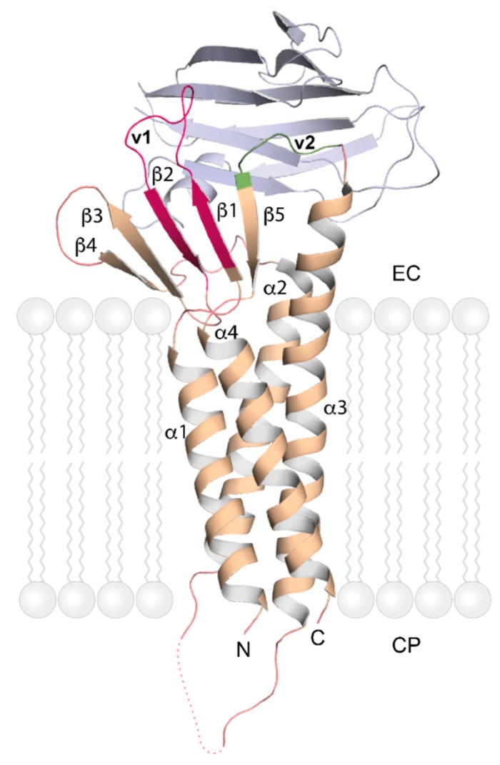
Figure 4.
Crystal structure of human claudin-4. Cartoon model of the overall fold of human CLDN-4 (wheat color) in complex with the C-terminal fragment of Clostridium perfringens enterotoxin (C-CPE, light blue; PDB entry 5B2G) [16,42]. The extracellular variable regions of CLDN-4 that mediate hetero- and homotypic interactions are highlighted in magenta (v1, comprising β1 and β2) and green (v2, between TM-helix α3 and β5), respectively. The dotted line marks a segment of polypeptide chain not represented in electron density. The stylized lipid molecules indicate the cell membrane and are not part of the experimental structure. EC: Extracellular; CP: Cytoplasmic.

