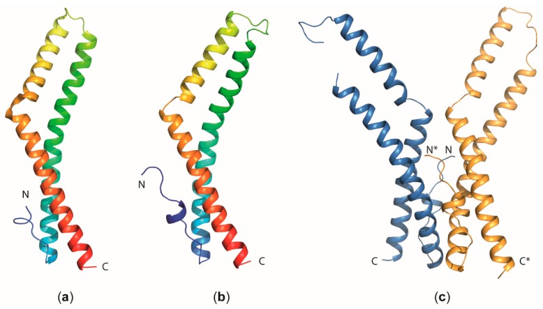
Figure 5.
Structural insight into occludin and tricellulin function. Cartoon models of the overall fold of the coiled-coil domain of (a) human occludin (PDB entry 1XAW) [58] and (b) human tricellulin (PDB entry 5N7K) [71]. The molecules are colored in a gradient ranging from blue at the N-terminus (N) to red at the C-terminus. (c) Dimeric arrangement of the tricellulin C-terminal coiled-coil domain observed in the crystal structure [71]. The chain marked with an asterisk (*) corresponds to the second monomer within the dimer.

