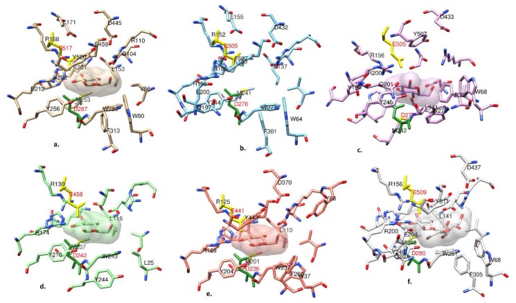
Figure 6.
Active site structure comparisons of (a) CtBGL, (b) NcCel3A, (c) ReCel3A, (d) TnBgl3B, (e) HjCel3A, and (f) AaBGL1. The corresponding conserved nucleophile Asp is colored in dark green and the acid/base Glu is colored in yellow. Both residues are labeled in red. Carbon atoms are colored differently in each enzyme. The active site BGC is displayed with its solvent-excluded surface for visual clarity.

