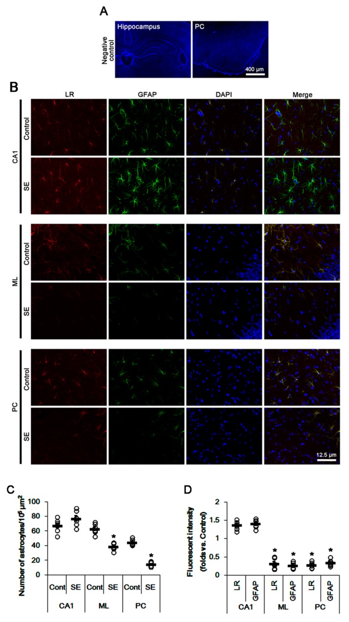
Figure 2.
Expressions of 67-kDa LR (LR) and glial fibrillary acidic protein (GFAP, an astroglial maker) in the hippocampus and the PC at 3 days after SE. Reactive astrocytes showed strong 67-kDa LR expression in the CA1 region at 3 days after SE. However, 67-kDa LR expression was reduced in the molecular layer of the dentate gyrus (ML) and the PC due to SE-induced astroglial degenerations. (A) Representative photographs of negative control of 67-kDa LR in the hippocampus and the PC (DAPI counterstaining). (B) Representative photographs of 67-kDa LR and GFAP in the hippocampus and the PC. (C,D) Quantitative values (mean ± S.E.M) of the number of astrocytes (C) and the fluorescent intensities of 67-kDa LR and GFAP (D) at 3 days after SE (n = 7, respectively). Open circles indicate each value. Horizontal bars indicate the mean value. Significant differences are * p < 0.05 vs. control animals (unpaired Student’s t-test).

