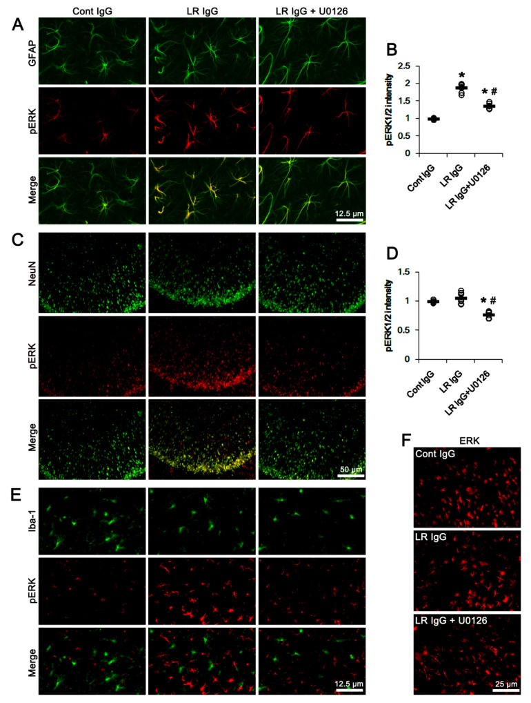
Figure 10.
Effects of 67-kDa LR (LR) neutralization on ERK1/2 and pERK1/2 expression in the PC of normal animals. Similar to the hippocampus, 67-kDa LR IgG infusion elevated pERK1/2 level in astrocytes, but not CA1 neurons and microglia, without changing ERK1/2 expression. U0126 attenuated pERK1/2, but not ERK1/2, levels in these cells following 67-kDa LR IgG infusion. (A) Representative photographs for pERK1/2 level in the PC astrocytes. (B) Quantitative values (mean ± S.E.M) of pERK1/2 level in the PC astrocytes (n = 7, respectively). Open circles indicate each value. Horizontal bars indicate the mean value. Significant differences are *,# p < 0.05 vs. control IgG and 67-kDa LR IgG, respectively (one-way ANOVA followed by Newman–Keuls posthoc test). (C) Representative photographs for pERK1/2 level in the PC neurons (NeuN, a neuronal marker). (D) Quantitative values (mean ± S.E.M) of pERK1/2 level in PC neurons (n = 7, respectively). Open circles indicate each value. Horizontal bars indicate the mean value. Significant differences are *,# p < 0.05 vs. control IgG and 67-kDa LR IgG, respectively (one-way ANOVA followed by Newman–Keuls posthoc test). (E) Representative photographs for pERK1/2 level in the PC microglia (Iba-1, a microglia marker). (F) Representative photographs for ERK1/2 level in the PC.

