Figure 12.
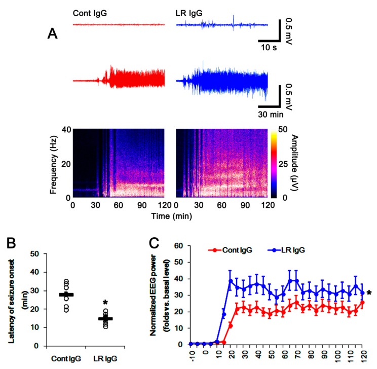
Effects of control IgG and 67-kDa LR IgG on seizure activity in response to pilocarpine. As compared to control IgG infusion, 67-kDa LR neutralization induced paroxysmal discharges on the baseline EEG in the hippocampus 3 days after infusion. In addition, 67-kDa LR IgG reduced seizure latency and increased seizure severity in response to pilocarpine. The 67-kDa LR IgG infusion reduced seizure latency and increased seizure severity in response to pilocarpine. (A) Representative baseline EEG (upper traces), seizure activity (lower traces), and frequency-power spectral temporal maps in response to pilocarpine. (B) Quantification of latency of seizure onset in response to pilocarpine (mean ± S.E.M.; n = 7, respectively). Open circles indicate each value. Horizontal bars indicate the mean value. Significant differences are * p < 0.05 vs. control IgG (unpaired Student’s t-test). (C) Quantification of total EEG power (seizure intensity) in response to pilocarpine (mean ± S.E.M.; n = 7, respectively). Significant differences are * p < 0.05 vs. control IgG (one-way repeated measure ANOVA). EEG: electroencephalogram.

