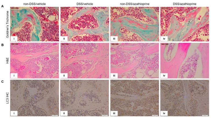
Figure 4.
Histological staining and immmunohistochemical labelling of tibia trabecular bone in sections of the tibia. (A) Goldner’s Trichrome, (B) H&E, (C) LC3 immunolabelling. LC3-positive immunolabelling is presented as brown staining. (i) non-DSS/vehicle; (ii) DSS/vehicle; (iii) non-DSS/azathioprine; (iv) DSS/azathioprine. Scale bar = 50 µm. Images are representative of 4 different mice/group.

