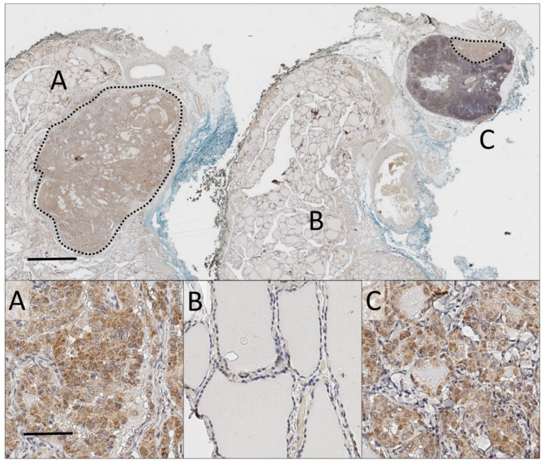
Figure 2.
Whole-slide analysis of a primary thyroid cancer and corresponding thyroid cancer nodal metastasis. Main: Low magnification (0.5×) of whole-slide of proNGF immunostaining (DAB, brown), with haematoxylin nuclear counterstain (blue), of primary tumour and paired metastasis. Blue and black inking represent the anterior and tracheal borders of thyroid respectively. Scale bar 2mm. (A) 7 mm microPTC with staining for proNGF. (B) Adjacent normal thyroid tissue. (C) 2 mm lymph node micro-metastasis at upper edge of node, included in same anatomical block. Dotted lines demarcate tumour. Inset: 20× magnification of representative fields. Scale bar 50 µm.

