Figure 1.
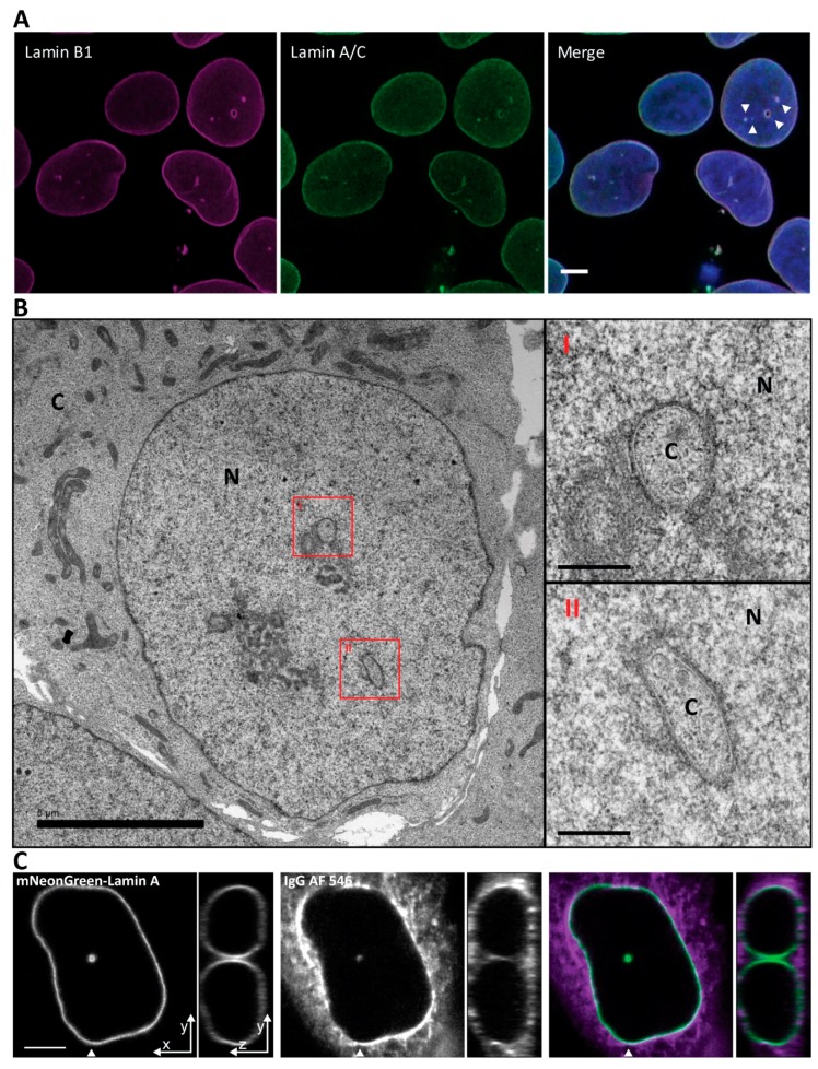
Nuclei of Ishikawa cells contain invaginations of the nuclear envelope forming a nucleoplasmic reticulum. (A) Ishikawa cells immunostained with anti-Lamin B1 and anti Lamin A/C. Scale bar 5 µm. (B) Electron microscopy micrograph of a high-pressure frozen Ishikawa cell. Scale bar is 5 µm for low magnification nucleus and 0.5 µm for high magnification insets. N, nucleus; C, cytoplasm. (C) Cytoplasmic loading by IgG AF 546 to visualize the cytoplasmic core of the invagination. Arrowhead indicates position of orthogonal projection in the panels to the right. Scale bar 5 µm.

