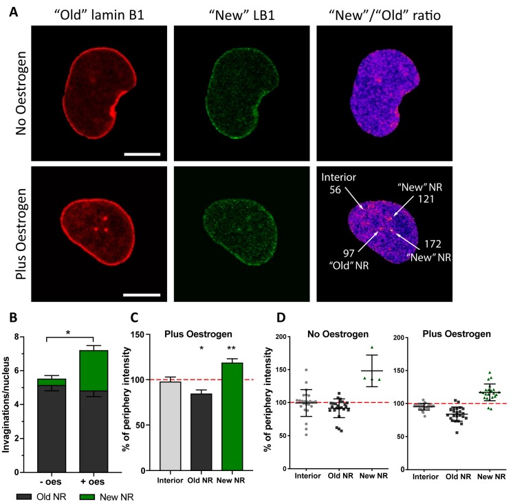
Figure 4.
Nascent lamin B1 is incorporated in newly formed invaginations. (A) Confocal microscopy of Ishikawa cells expressing lamin B1- Maple3. Indicated are the “old” (red channel) and “new” (green channel) lamin protein pools. Ratiometric image of “New”/”Old” is provided with indication of ratio values for selected ROIs around the features arrowed. (B) Evaluation of invagination abundance per nucleus in Ishikawa cells with (+ oes) or without oestrogen (-oes) treatment. (C) Pixel intensities of the ROIs defined in based on the ratiometric images and normalised to the signal at the nuclear rim showing increased incorporation of nascent lamin B1 at the newly forming NR channels; results from three independent experiments, 35 cells in total; mean ± SD; ** p-value < 0.001; * p-value < 0.05. (D) An example data plot from a single experiment showing distribution of “New”/”Old” lamin B1 ratio at different nuclear structures and normalised to the nuclear rim ratio with or without oestrogen.

