Figure 4.
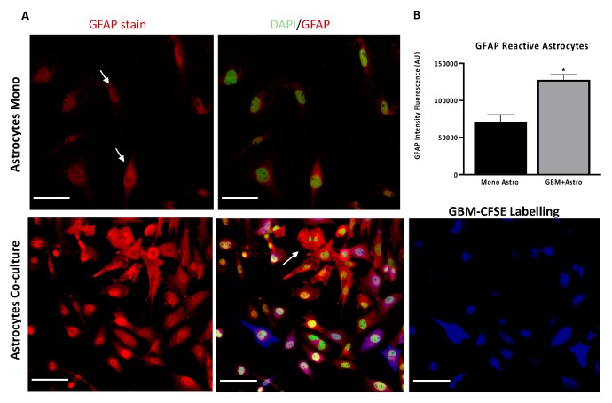
Human astrocytic cells become “reactive” in co-culture at 50:50 ratio with glioblastoma cells after 48 h. (A) Astrocytes co-cultured with GBM cells exhibited morphological changes and became ‘reactive’. Immunofluorescence staining for glial fibrillary acidic protein (GFAP) (Red) in normal astrocytes (UP-010) cultured alone or co-cultured with GBM (UP-007) cells. UP-007 cells were labelled with carboxyfluorescein succinimidyl ester (CFSE) (blue), and the nuclei were stained with Hoechst (in green). (B) Bar plot of mean fluorescence intensity of GFAP expression in astrocytes in co-culture or mono- culture. * p < 0.05 represents the statistical significance in a two-tailed Student’s t-test. Mean ± SEM (n = 2). Scale bar: 80 µm.

